The importance of diet in derm health
A thorough feeding history and a standard dermatologic exam can prevent missed diagnoses and place nutrition where it belongs: as a routine component of dermatology care for both healthy and allergic pets.
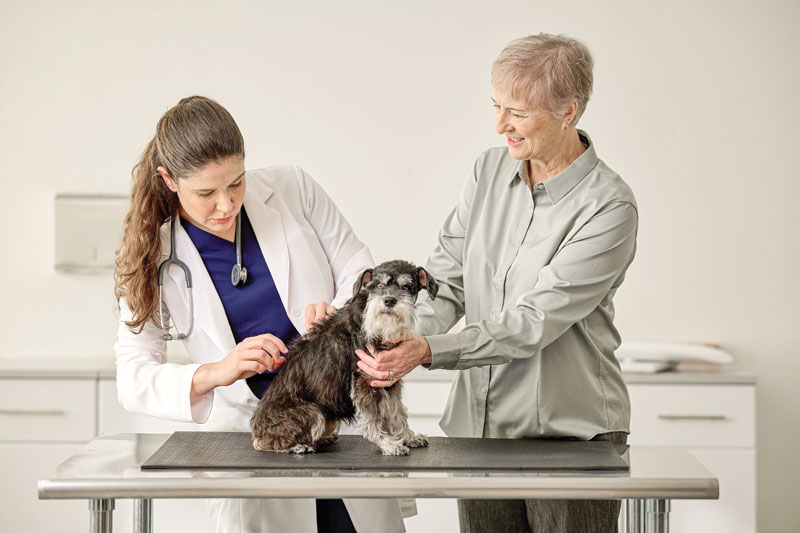
Skin and coat quality are among the most visible reflections of a pet’s overall health.1 Pets may present for dull coat, scaling, increased shedding—but those observations often indicate deeper issues in nutrition, barrier function, or early allergic skin disease. A thorough dietary history, coupled with a standard dermatologic exam, can prevent missed diagnoses and place nutrition where it belongs: as a routine component of dermatology care for both healthy and allergic pets.
Taking a complete history, including a diet history at every visit—well and sick alike—helps clinicians identify problems early and tailor the plan.
The nutritional foundation of healthy skin
Fatty acids: Building the barrier
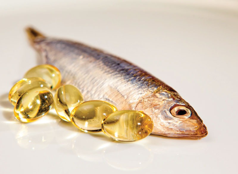
Essential fatty acids (EFAs) are cornerstone nutrients for cutaneous health. Linoleic acid (LA; omega-6) supports ceramide synthesis in the stratum corneum; adequate intake improves skin hydration and coat gloss and reduces transepidermal water loss (TEWL)—a measurable marker of skin barrier competence in healthy dogs.2
Long-chain omega-3 polyunsaturated fatty acids (PUFAs) (eicosapentaenoic acid [EPA], docosahexaenoic acid [DHA]), while not essential for the adult, modulate inflammatory pathways and stabilize membranes, contributing to skin homeostasis in healthy pets and serving as a valuable adjunct when inflammation is present. Because dogs—and especially cats— inefficiently convert precursors, such as alpha-linolenic acid (ALA) preformed EPA/DHA sources matter in practice.3
While true EFA deficiency is uncommon in complete and balanced diets, it can occur with unbalanced or unconventional feeding and conditions leading to malabsorption. Clinically, pets may present with dry, flaky skin, dull coat, seborrhea, alopecia, or increased TEWL; targeted dietary correction restores barrier function and coat quality.4,5
Protein and amino acids: Architecture for hair and skin
Hair is composed of approximately 95 percent protein; meeting amino acid needs is necessary for normal keratinization, pigmentation, and follicular cycling.1 Methionine and cysteine (sulfur amino acids) are heavily incorporated into hair; phenylalanine and tyrosine drive melanin synthesis.4 Protein shortfalls or amino acid imbalances can manifest as a brittle coat, altered color, or slowed growth.1 Most complete and balanced commercial diets provide adequate protein for healthy skin and hair, but a diet history is crucial to assess intake and catch at-risk pets.
Zinc, vitamin A, vitamin E: Micronutrients with macro effects
Zinc is critical for keratinocyte turnover, fatty acid metabolism, immune function, and wound healing; ~20 percent of total body zinc resides in skin, especially in keratinized tissues, including nose, paw pads, and tongue.6 Deficiency can produce parakeratosis, scaling, and a lackluster coat. Certain northern breeds, including Siberian huskies, Alaskan malamutes, and Samoyeds, are predisposed to zinc-responsive dermatosis4—making zinc bioavailability an important selection criterion when choosing diets or supplements.7 More bioavailable forms of zinc include zinc sulfate, gluconate, and methionine.
Vitamin A regulates follicular turnover and epithelial differentiation;8 both deficiency and chronic over-supplementation are dermatologically detrimental (e.g., follicular hyperkeratosis with deficiency; dry, flaky skin and alopecia with excess).9
Vitamin E plays vital roles in the skin and immune system. It is essential to protect PUFA-rich membranes, and it may need to increase when total dietary PUFA rises (e.g., fish oil–enriched diets or high-fat diets).10
Clinical takeaway: For well pets, most “coat problems” resolve with complete and balanced feeding and consistent owner adherence; supplementation is case-by-case and should be evidence-based.
Nutrition as a therapeutic adjunct in atopic dermatitis
Atopic dermatitis (AD) is a genetically influenced, inflammatory, and pruritic skin disease triggered by environmental allergens.11 Nutrition can help modify disease expression by strengthening the skin barrier, tempering inflammation, and supporting immune balance. Studies in dogs demonstrate that EFA-enriched diets, and in some cases, diets fortified with antioxidants/polyphenols, can reduce pruritus scores and lesion indices and decrease reliance on concurrent antipruritics, albeit with variable effect sizes across trials.12, 13
Palmitoylethanolamide (PEA), an endocannabinoid-like lipid, has been shown to reduce pruritus and improve remission maintenance in both dogs and cats when used as a component of multimodal therapy.14,15
The evidence base for atopy supported by nutrition is varied due to a number of assessment methods, including CADESI, CADLI, SCORFAD, and PVAS, that score skin lesions and pruritus, but a pattern emerges: EFA-enriched nutrition and barrier-supportive formulations can provide clinically meaningful additive benefits over time. For client communication, frame nutrition as a “stabilizer” that helps reduce flare intensity and extend comfortable intervals—not a cure.
Cutaneous adverse food reaction (CAFR): Diet, friend or foe?
Cutaneous adverse food reactions can be indistinguishable clinically from atopy and frequently coexist with it.16 Dogs and cats with CAFR may present with non seasonal pruritus, recurrent otitis, secondary infections, and GI signs (vomiting, diarrhea, increased bowel movements, gas, borborygmus).17 Because there is substantial overlap with atopy, diagnosis depends on a strict elimination diet trial (See: “The elimination diet: Practical, clinic-ready steps”) followed by a dietary rechallenge—serologic, saliva, hair, intradermal, or patch tests are not reliable for confirming food allergy.18
Practical communication strategies that work:
Putting it all together in general practice
|
Communication with owners: The determining factor
Across nutrition and dermatology references, the strongest predictor of success is not the protein source or degree of hydrolysis—it is owner understanding and adherence. Diet trials commonly fail due to small, repeated “leaks:” neighbors giving treats, flavored preventatives or medications, pill hiding in cheese, multi-pet food sharing, dropping food, or scavenging. Bringing these pitfalls into the open normalizes the challenge and prevents unintentional noncompliance (See: “Practical communication strategies that work”).
The elimination diet: 6 Practical, clinic-ready steps
Timelines at a glance (for client education):
|
Advances in dermatologic health
Persistent skin issues in companion animals go beyond surface-level concerns. They arise from complex interactions among the skin barrier, immune system, and cutaneous microbiome. Research shows when the skin’s protective barrier is disrupted, sensory signals linked to discomfort intensify, fueling an itch–scratch cycle that further weakens skin integrity. Subclinical inflammation and shifts in barrier lipid composition are now recognized as early contributors, often appearing before visible lesions develop. These findings highlight the importance of proactive skin support strategies that strengthen barrier resilience and help calm underlying inflammation, rather than relying solely on symptomatic relief once clinical signs become more pronounced.
Conclusion
Nutrition is inseparable from dermatologic health. In healthy pets, truly complete and balanced diets provide the substrates—EFAs, amino acids, zinc, vitamins A and E—that sustain barrier function and coat vitality. In atopic pets, nutrition adds measurable value by supporting the epidermal barrier and tempering inflammation as part of a multimodal plan. In food allergy, diet serves as both a diagnostic tool and a long-term nutritional management tool, provided trials are conducted strictly and confirmed by rechallenge.
With clear, empathetic communication and a structured approach, veterinarians can turn complex skin cases into manageable, evidence-based successes that improve comfort and strengthen the veterinarian–client relationship.
Jason Gagné, DVM, DACVIM (Nutrition), is Purina Institute’s director of External Relations. Prior to his role at the company, and throughout his residency at Cornell, Dr. Gagné served as an associate veterinarian in a small animal practice in Syracuse, N.Y. Gagné has authored several publications in veterinary journals and textbooks, given scientific presentations at the regional, national, and global level, and serves as a scientific reviewer for several journals.
References
- Scott, D.W., Miller, W.H., & Griffin, C.E. (2001), Muller & Kirk’s Small Animal Dermatology (6th edition). Saunders.
- National Research Council (NRC). (2006). Nutrient Requirements of Dogs and Cats. The National Acadamies Press.
- Bauer JE. Therapeutic use of fish oils in companion animals. J Am Vet Med Assoc. 2011 Dec 1;239(11):1441-51.
- Outerbridge, C.A. & Owens, T.J. (2023). Nutritional Management of Skin Diseases. In A.J. Fascetti, S.J. Delaney, J.A. Larsen, & C. Villaverde (Eds.), Applied Veterinary Clinical Nutrition (2nd, pp. 345-383). Wiley-Blackwell.
- Watson, A., Thomas, G., Butkowski, C., & Allaway, D. (2018). Evidence for an interaction between linoleic acid intake and skin barrier properties in healthy dogs – A pilot study. Journal of Applied Animal Nutrition, 6 e7.
- Roudebush P, Wedekind KJ. Zinc-responsive dermatosis in dogs. Vet Dermatol. 2002 Feb;13(1):63.
- Logas, D., Kunkle, G.A., & McDowell, L. (1993). Comparison of Serum Zinc Levels in Healthy, Systemically Ill and Dermatologically Diseased Dogs. Veterinary Dermatology,4(2), 61-64.
- Dodd S, Cave N, Abood S, Shoveller AK, Adolphe J, Verbrugghe A. An observational study of pet feeding practices and how these have changed between 2008 and 2018. Vet Rec. 2020 Jun 27;186(19):643
- Case, L.P., Daristotle, L., Hayek, M.G., & Raasch, M.F. (2011). Canine and Feline Nutrition (3rd). Elsevier.
- Association of American Feed Control Officials. (2023). AAFCO Dog and Cat Nutrient Profiles.
- Halliwell R. Revised nomenclature for veterinary allergy. Vet Immunol Immunopathol. 2006 Dec 15;114(3-4):207-8.
- Logas D, Kunkle GA. Double-blinded Crossover Study with Marine Oil Supplementation Containing High-dose icosapentaenoic Acid for the Treatment of Canine Pruritic Skin Disease. Vet Dermatol. 1994 Sep;5(3):99-104.
- Scarff DH, Lloyd DH. Double blind, placebo-controlled, crossover study of evening primrose oil in the treatment of canine atopy. Vet Rec. 1992 Aug 1;131(5):97-9.
- Noli C, Della Valle MF, Miolo A, Medori C, Schievano C; Skinalia Clinical Research Group. Efficacy of ultra-micronized palmitoylethanolamide in canine atopic dermatitis: an open-label multi-centre study. Vet Dermatol. 2015 Dec;26(6):432-40, e101.
- Noli C, Della Valle MF, Miolo A, Medori C, Schievano C; Skinalia Clinical Research Group. Effect of dietary supplementation with ultramicronized palmitoylethanolamide in maintaining remission in cats with nonflea hypersensitivity dermatitis: a double-blind, multicentre, randomized, placebo-controlled study. Vet Dermatol. 2019 Oct;30(5):387-e117.
- Hillier A, Griffin CE. The ACVD task force on canine atopic dermatitis (X): is there a relationship between canine atopic dermatitis and cutaneous adverse food reactions? Vet Immunol Immunopathol. 2001 Sep 20;81(3-4):227-31.
- Gaschen, F. P., Merchant, S. R. (2011). Adverse food reactions in dogs and cats. Vet Clin North Am Small Anim Pract, 41(2), 361-379.
- Mueller RS, Olivry T. Critically appraised topic on adverse food reactions of companion animals (4): can we diagnose adverse food reactions in dogs and cats with in vivo or in vitro tests? BMC Vet Res 2017;13(1):275.
- Rybnícek J, Lau-Gillard PJ, Harvey R, et al. Further validation of a pruritus severity scale for use in dogs. Vet Dermatol. 2009 Apr;20(2):115-122.
- Olivry T, Saridomichelakis M, Nuttall T, et al. International Committee on Allergic Diseases of Animals (ICADA). Validation of the Canine Atopic Dermatitis Extent and Severity Index (CADESI)-4, a simplified severity scale for assessing skin lesions of atopic dermatitis in dogs. Vet Dermatol. 2014 Apr;25(2):77-85, e25

Sign up for our newsletter
Get all the latest news and features from Veterinary Practice News Canada. Submit your email below to get our twice-monthly newsletter.
Popular Articles
-
1
-
2
-
3
-
4
-
5
Read the Latest Issue

