Hip Dysplasia: Classic Changes On X-rays
Changes regarding the reading of X-rays of hip displaysia.
When evaluating dysplastic hip films, radiographic changes may include:
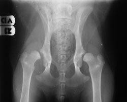
A 7-month-old male Labrador suffering from severe hip dysplasia.
• Hip subluxation, i.e. less than 66 percent coverage of the femoral head by the acetabulum.
• The margins of the acetabulum and femoral head are not parallel. They form a triangle or a wedge.
• Increased width of the joint space.
• Thickening of the femoral neck.
• Flattening or deformity of the femoral head.
• Flattening of the acetabulum.
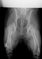
A 6-year-old female Rottweiler suffering from severe hip dysplasia.
• Irregular acetabulum rim.
• Osteophytes on the acetabulum, femoral head and neck.
• Sclerosis of the subchondral bone.
A description of a normal hip would include:
• Two-thirds of the femoral head are covered by the acetabulum.
• The margins of the acetabulum and femoral head are parallel.
• A small, flattened area of the femoral head represents the fovea capitis, which is where the round ligament attaches. This is a normal finding.

Sign up for our newsletter
Get all the latest news and features from Veterinary Practice News Canada. Submit your email below to get our twice-monthly newsletter.
Read the Latest Issue

