Is LLLT over active epiphyses contraindicated?
Whichever course of action is best for an individual patient must take priority
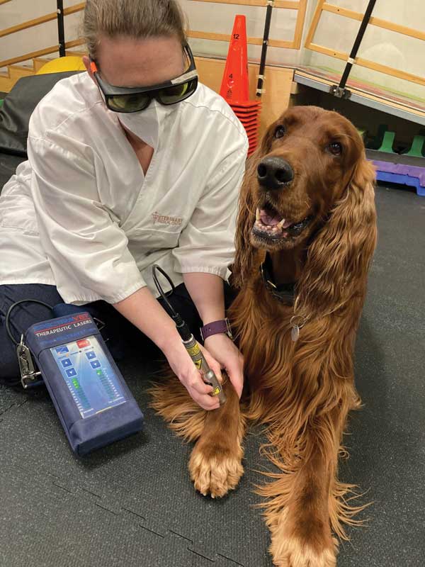
Photo courtesy Kira Penney
An often-recurring question regarding the clinical use of laser therapy is whether treating over active epiphyses is contraindicated.
A general list of contraindications to low-level laser therapy (LLLT)—or, formally, photobiomodulation (PBM)—in the treatment of musculoskeletal disorders has largely been carried over from other active modalities, such as ultrasound and electrical stimulation (e-stim), and has been accepted without question (with, of course, the same abundance of caution as applying to laser therapy in general).
The use of laser over or near the active epiphysis seemed, initially, to have fit this category. It is often mentioned in device-operating manuals and by clinical educators in various fields as being contraindicated, but with little to no explanation as to why; however, others have considered this use to be of no great concern (again, with no explanation).
This article offers a brief review of relevant literature while seeking to answer the question: Is LLLT/PBM over active epiphyses truly contraindicated?
Research
In 2010, the Canadian Physiotherapy Association (CPA) published, ‘Electrophysical Agents Contraindications and Precautions: An Evidence-Based Approach to Clinical Decision Making in Physical Therapy.’1 According to this document, LLLT/non-coherent light is considered safe for use “on skin overlying active epiphysis,” and “can be applied with caution” to the active epiphysis.
To this author’s knowledge, this is the first (and, possibly, only) formal guidance document of this type to address the safety of LLLT/PBM in an evidence-based manner. Further, its recommendations for physiotherapists were contemporaneously accurate and well-founded—but, more than a decade after being published, is it still current?
On a hunt for answers, I conducted a search of PubMed, Google Scholar, ResearchGate, and a number of laser- and PBM-related journals and texts using various combinations of key words, including:
- laser therapy
- LLLT
- photobiomodulation
- PBM
- epiphysis
- epiphyses
- epiphyseal; and
- growth plate.
This query turned up relatively few relevant articles,2-11 one indirectly relevant paper,12 and a number of false positives—two in the latter category13,14 are often inappropriately cited in lay discussion as ‘evidence’ of the negative impact of laser therapy on active epiphyses.
When the CPA document was published in 2010, the only readily available literature on the subject was a study by Cheetham et al (1992),2 which investigated the effects of laser therapy over active epiphyses. In 2002, Navratil et al3 referred to this when stating, “the fear of possible damage to the epiphyseal slots in children in the case of the application of therapeutic laser is baseless.” Other work4,5 had also been done at the time, but accessibility to the literature is limited, as it has only ever been published in Russian.
Since 2010, six more directly relevant papers6-11 have been published, along with one more12 containing pertinent information, albeit from within a different field. The various studies shed more light on the effects of LLLT/PBM on epiphyseal growth, but the results remain somewhat controversial.
For example, de Andrade8 and Handayani11 found no significant effect on the histology of the epiphyseal cartilage or the final length of limbs, but Cressoni7 demonstrated laser irradiation may improve cartilage structure in rats. Meanwhile, Seifi,6 Oliveira,9 and Yeom10 all found significant histological and histomorphometric changes, particularly within the hypertrophic zone, as well as changes in bone length. Oliveira9 found the femoral longitudinal length decreased, whereas Yeom10 found an increased rate of growth. Finally, while investigating the impact of photodynamic therapy upon epiphyseal plates, Kurchenko12 found laser irradiation without the introduction of a photosensitizer led to intracellular swelling of epiphyseal plates chondrocytes.
Considerations
A major confounding factor when considering these findings in relation to our initial question is that each of these studies was conducted with widely differing device and treatment parameters. What’s more, the devices used bear scant resemblance to the majority of those currently in clinical use.
Where reported, the ‘stationary contact’ application technique was consistent among all studies, but the number of points irradiated, as well as the number of irradiations performed over time, varied greatly. Indeed, output powers ranged from 4 to 100 mW, power densities from 2 to 563 mW/cm2, irradiation durations from 8.5 seconds to eight minutes, wavelengths from 635 to 904 nm, and dosimetry was all over the shop. (To view Table 1, click here.)
Essentially, there is little basis for meaningful comparison of these studies, other than they were investigating effects upon similar tissues by applying laser light to similar anatomical locations.
Two studies10,11 treated acupuncture points rather than specific anatomical locations over the growth plates; however, two of the points used (ST36 and SP6) are located adjacent to growth plates—ST36 about a millimetre lateral of the tibial tuberosity, and SP6 a few millimetres above the medial malleolus on the posterior border of the medial aspect of the tibia. Perhaps unsurprisingly, irradiation over these points elicited greater changes than irradiation of GV20, which is located at the peak of the head, midpoint of a line connecting the apexes of the two auricles (Figure 1).
Although some studies found no effects of laser irradiation at the parameters used, upon active epiphyses in rats and hamsters, others did. Thus, there is certainly evidence of effects, including increased angiogenesis, increased proliferation of chondrocytes, and increased calcification.
Not all of the potential effects of LLLT/PBM on growth plates are, necessarily, negative. Yeom et al,10 for example, conclude longitudinal bone growth induced by laser acupuncture to points directly over growth plates, “may have a clinical potential in promoting longitudinal bone growth in children,” and Seifi et al6 suggest, “laser irradiation with the chosen parameters can stimulate condylar growth and subsequently cause mandibular advancement […] for further improvement of mandibular retrognathism.” Finally, according to Mavrich and Luzin,4 laser can be used for the optimization of growth, mineralization, and stability of skeleton bones.

Image courtesy SpectraVET
Uncertainties remain
So, where does this leave us?
With more information, we can make better-informed clinical choices; however, with the current data, it is not possible to determine an upper- or lower-margin for any of the important parameters, nor to accurately define a ‘window’ of effect. It is worth noting, too, no studies have yet considered the effect of high-powered (i.e. Class 4 or IV) ‘therapeutic’ lasers and/or very high doses.
If one does not look too closely at the weeds and seeds, it might be possible to find a ‘big picture’ pattern among the data suggesting: (a) more frequent treatments with higher intensities and/or doses over longer periods are more likely to have deleterious effects than (b) less-frequent treatments with lower intensities/doses over shorter periods.
Even with these new data, though, the only thing apparent is there is still no clear answer to the initial question posed. There is evidence of effects of LLLT/PBM upon active epiphyses, and, in some cases, this may prove to be strongly contraindicated. What is not addressed by any these studies, however, is the relative benefit or otherwise of utilizing LLLT/PBM in young patients versus withholding treatment due to concerns over the impact to growth plates.
Whichever course of action is best for an individual patient must take priority; there will no doubt be many instances where, despite any possible risks, LLLT/PBM is strongly indicated. In all cases, however, a cautious and well-considered approach is recommended.
Peter Jenkins [ORCID ID: 0000-0002-8456-5919] is founder and director of education and technology for SpectraVET Inc., a U.S.-based developer and manufacturer of veterinary laser/LED/IRED photobiomodulation (PBM) therapy devices. He also co-founded the Australian Medical Laser Association (now Australian Medical Photobiomodulation Association– AMPA) and Immunophotonics Inc., a biotech company focused on cancer immunotherapy.
References
1 Physiotherapy Canada (2010) ELECTROPHYSICAL AGENTS Contraindications and Precautions: An Evidence-Based Approach to Clinical Decision Making in Physical Therapy V62/5 Special Issue.
2 Cheetham MJ, Young SR, and Dyson M. (1992) Histological effects of 820 nm laser irradiation on the healthy growth plate of the rat. Laser Therapy, vol. 4, no. 2, pp. 59-63.
3 Navrátil L and Kymplova J. (2002) Contraindications in noninvasive laser therapy: truth and fiction. J. Clin. Laser Med. Surg. 20, 341-343.
4 Mavrich VV, Luzin VI. (2000) The growth, chemical composition and strength properties of the long tubular bones in the skeleton of white rats under the influence of low energy laser radiation. Morfologiia. 2000;117(1):59-66. [Article in Russian]
5 Pereslytskikh PF (2008) Formation of ossification nucleus in the femoral head in hamsters exposed to laser radiation. Morfologiia. 2008;134(6):68-72. [Article in Russian]
6 Seifi M, Maghzi A, Gutknecht N, Mir M, Asna-Ashari M. (2010) The effect of 904 nm low level laser on condylar growth in rats. Lasers Med Sci. 2010;25(1):61-5.
7 Yeom M, Kim S-H, Lee B, Zhang X, Lee H, Hahm D-H, Sohn Y, Lee H. (2013) Effects of Laser Acupuncture on Longitudinal Bone Growth in Adolescent Rats. Evidence-Based Complementary and Alternative Medicine: eCAM, 2013, 424587.
8 Cressoni MDC, Giusti HHKD, Piao AC, de Paiva Carvalho RL, Anaruma CA and Casarotto RA. (2010) Effect of GaAlAs laser irradiation on the epiphyseal cartilage of rats. Photomedicine and laser Surgery, 28(4), pp.527-532.
9 de Andrade AR, Meireles A, Artifon EL, Brancalhão RMC, Ferreira JRL and Bertolini GRF. (2012) The effects of low-level laser therapy, 670 nm, on epiphyseal growth in rats. The Scientific World Journal, 2012.
10 Oliveira SP, Rahal SC, Pereira EJ, Bersano PRD, Vieira FD, and Padovani CR. (2012) Low-level laser on femoral growth plate in rats. Acta Cirurgica Brasileira, vol. 27, no. 2, pp. 117-122, 2012.
11 Handayani S, Ramelan AH, Purwanto B, Saputra K and Tamtomo DG (2017) Histology of Epiphyseal Plate of Adolescent Rat Stimulated by Laserpuncture. J. Phys.: Conf. Ser. 909 012092
12 Kurchenko S, Shashko A, Dudin M, Mikhailov V, Netylko G, Ashmarov V. (2012) Photodynamic impact on the epiphyseal plates. Stud Health Technol Inform. 2012;176:174-8.
13 Morein G, Gassnerb S, and Kplanc I. (1978) Bone growth alterations resulting from application of CO2 laser beam to the epiphyseal growth plates: an experimental study in rabbits. Acta Orthopaedica Scandinavica, vol. 49, no. 3, pp. 244-248, 1978.
14 Peterson HA and Wood MB. (2001) Physeal arrest due to laser beam damage in a growing child. J. Pediatr. Orthop. 21, 335-337.
Sign up for our newsletter
Get all the latest news and features from Veterinary Practice News Canada. Submit your email below to get our twice-monthly newsletter.
Popular Articles
-
1
-
2
-
3
-
4
-
5
Read the Latest Issue
