
Reviewing best treatment options for periodontal disease
Making the diagnosis of periodontal disease is not typically a challenge. The challenge is quantifying each tooth’s disease severity and understanding the options for treating each of them individually.
Life is straightforward, albeit boring, when medical decision-making is “cut and dried.” Healthy teeth? Clean and polish. A severely diseased tooth? Extract. However, as in every aspect of life, there are shades of grey when making decisions in treating periodontal disease.
Making the diagnosis of periodontal disease is not typically a challenge. The challenge is quantifying the disease severity of each tooth and understanding the options for treating each individually. In dogs and cats, periodontal disease is typically a slowly progressive condition that may present at varying stages of severity. Table 1 below shows the staging nomenclature used by the American Veterinary Dental College.1
| PD 0 (Normal) | Clinically normal; no gingival inflammation or periodontitis clinically evident. |
| PD 1 (Stage 1) | Gingivitis only; without attachment loss. The height and architecture of the alveolar margin are normal. |
| PD 2 (Stage 2) | Early periodontitis, with less than 25 percent of attachment loss, or at most, there is a stage 1 furcation involvement in multi-rooted teeth. There are early radiologic signs of periodontitis. The loss of periodontal attachment is less than 25 percent as measured either by probing of the clinical attachment level or radiographic determination of the distance of the alveolar margin from the cementoenamel junction relative to the length of the root. |
| PD 3 (Stage 3) | Moderate periodontitis, with 25-50 percent of attachment loss as measured either by probing of the clinical attachment level, radiographic determination of the distance of the alveolar margin from the cementoenamel junction relative to the length of the root, or stage 2 furcation involvement in multi-rooted teeth. |
| PD 4 (Stage 4) | Advanced periodontitis, with more than 50 percent of attachment loss as measured either by probing of the clinical attachment level, or radiographic determination of the distance of the alveolar margin from the cementoenamel junction relative to the length of the root, or there is stage 3 furcation involvement in multi-rooted teeth. |
When I teach students, I tell them there are 42 patients in a dog’s mouth and 30 in a cat’s mouth. Each “patient” requires an assessment and a plan, and documented justification for the recommended plan.
Types of periodontal bone loss
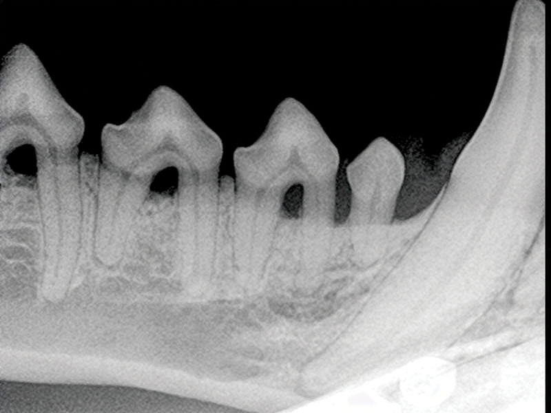
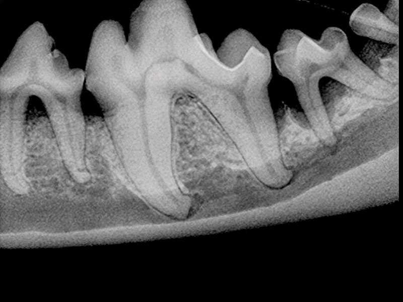
Periodontal bone loss can be divided into two categories: horizontal and vertical. Horizontal bone loss refers to alveolar bone loss that occurs along the alveolar margin of the mandible or maxilla, resulting in root exposure and furcation exposure of multi-rooted teeth (Figure 1). Vertical bone loss is alveolar bone loss that occurs along the long axis of a root (Figure 2). These two processes may occur simultaneously around the alveolar bone of the same tooth, or even the same root. Of the two processes, vertical bone loss is easier to combat than horizontal bone loss, since there are often three walls of bone surrounding the vertical defect that can retain osteoconductive or osteoinductive substances.
When to recommend advanced therapy
Has the periodontal disease progressed to the point where endodontic disease is now also likely? If so, the “double whammy” of periodontal disease and endodontic disease makes extraction the only practical choice.
Before deciding whether to pursue periodontal therapy for borderline teeth, it is important to have a heart-to-heart conversation with the pet owner. Is the affected tooth functionally important based on the patient’s role in life (police dog or couch potato?) Is the owner concerned about tooth loss? Is the patient healthy enough to undergo future anesthetic follow-up and treatment, since advanced periodontal disease often requires multiple treatments? Is the owner able and willing to perform daily home care? If not, even the best periodontal surgeries may fail over time due to the progression of periodontal disease. Another factor to consider is if you have the equipment, materials, and experience to provide advanced periodontal therapy.
Tools of the trade
In next month’s column, I will discuss the growing variety of periodontal products/devices that can be used in periodontal pockets. However, it is important to know what you put into a periodontal defect is less important than what you remove from the pocket. Prior to placement of any product, it is very important to perform a thorough ultrasonic scaling with an ultrasonic tip designed for subgingival scaling. Next, hand curettes should be used to perform root planing to remove remaining calculus deposits from the root surface and to make it as smooth as possible. Next, the hand curette is used to perform subgingival curettage, where the cutting surface of the curette is used on the soft tissue of the walls of the pocket to remove diseased granulation tissue, resulting in freshly cut surface of gingival connective tissue to reattach to the root surface. The site is then irrigated to flush debris.
Such techniques are not for every patient or every pet owner, but good outcomes can be seen, especially in cases of vertical bone loss in the absence of concurrent endodontic disease and tooth mobility. Consider attending a wet lab on the techniques of advanced periodontal therapy before performing these procedures.
Recommended reading materials:
|
John R. Lewis, VMD, DAVDC, Fellow-AVDC OMFS, practices at Veterinary Dentistry Specialists and teaches at Silo Academy Education Center, both in Chadds Ford, Pa.
Reference

Sign up for our newsletter
Get all the latest news and features from Veterinary Practice News Canada. Submit your email below to get our twice-monthly newsletter.
Popular Articles
-
1
-
2
-
3
-
4
-
5
Read the Latest Issue

